Developmental Neuropathology
Chapter 6: Polymicrogyria
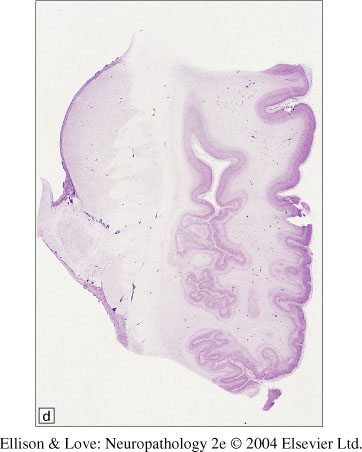
Polymicrogyria: cerebral cortical malformation with excessively folded cortical ribbon of miniature, individually thin convolutions, either fused together or piled on top of each other.
- increasingly recognized with MRI, no incidence data available
- some forms of polymicrogyria are more common in males
- risk factors include positive family history, intrauterine infection (CMV, toxoplasma, VZV, syphilis), intrauterine hypoperfusion (more commonly symmetrical MCA territory), loss of a twin/triplet
- associated with metabolic disorders, e.g., Zellweger syndrome
- associated with other congenital anomaly syndromes, e.g., 22q deletion, 1p36 deletion, Adams-Oliver syndrome, Niikawa-Kuroki syndrome
- also seen in Pelizaeus-Merzbacher, glutaric acidemia type II, thanatophoric dysplasia, maple syrup urine disease, histidinemia, Leigh sundrome, mitochondrial respiratory chain disorder, neonatal adrenoleukodystrophy
- autosomal dominant, recessive, and X-linked inheritance reported
- no specific genes have been identified, but polymicrogyria has been seen in a number of different mutations
Clinical features
- often undiagnosed until their child is diagnosed
- undiagnosed cases usually have localized polymicrogyria, and have medical or education histories
- most common childhood presentation is developmental delay, spastic tone, seizures, and microcephaly
- Worster-Drought syndrome - perisylvian polymicrogyria, associated with pseudobulbar palsy
- other clinical associations include sensorineural deafness, arthrogryposis, and other underlying syndromes
- can be diagnosed on MRI (apparent cortical thickening and irregular grey-white junction, often with reduction in white matter)
- radiological classification as unilateral/bilateral, predominantly frontal, perisylvian, parieto-occipital, or diffuse
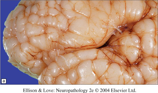
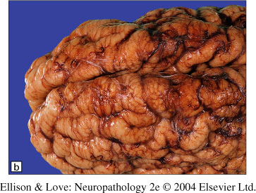
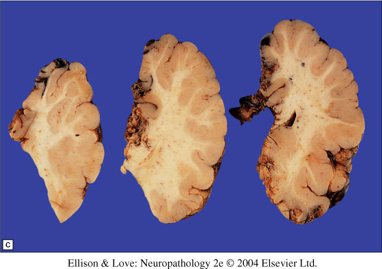
Macroscopy
- irregular, bumpy surface
- on coronal section, cortex is heaped up with fused mini-gyri producing the appearance of a thick cortical ribbon
- grey-white junction recognisable but disorganized
- can be widespread or limited to lobe or vascular territory
- often found bordering porencephaly or in the temporal lobes of hydranencephaly
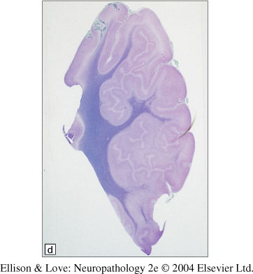
Histopathology
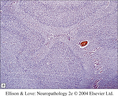
- thin, abnormally laminated, excessively folded cortical ribbon with fusions to adjacent ribbons
- two subtypes, which may occur together:
- unlayered polymicrogyria - thin unlayered band of grey matter interrupted by branching tissue from the molecular layer with a central blood vessel - fusing of adjacent molecular layers.
- 4-layered polymicrogyria - comprised of outer molecular layer, followed by two neuronal layers separated by a paucicellular band with myelinated fibers
- associated malformations include glioneuronal leptomeningeal heterotopia, nodular heterotopia, types I and II lissencephaly
Pathogenesis
- acquired or intrinsic malformations
- association with hypoperfusion around 3-5 months gestation, it coincides with neuronal migration
- also hypothesized to be due to post-migrational destruction of the cortex, since the 4 layered cortex corresponds architechurally to the first 4 layers of the 6 layered cortex (missing layers)